As the future of materials science is nano, Photo-induced Force Microscopy (PiFM) is an indispensable tool to analyze the topology and chemical makeup of new materials in exquisite detail.

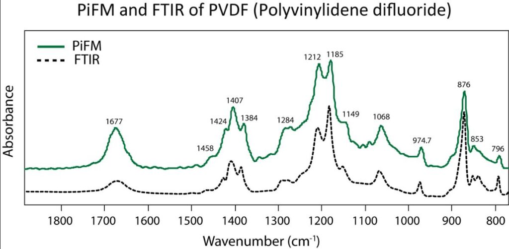
Photo-induced Force Microscopy (PiFM), combines infrared (IR) absorption spectroscopy with atomic force microscopy (AFM) which results in high spatial resolution chemical images and spectra. Photo-induced force microscopy measures the local absorption resonances in the sample by the mechanical detection of forces in the tip-sample junction. By mapping the IR absorption of the sample as a function of IR wavelength and position, nm-scale resolution reveals the locations of heterogeneous materials. Moreover, the IR laser can be tuned to generate spectral information that is like FTIR measurements with less than 10 nm spatial resolution as shown in the images above.
Photo-induced force microscopy measurements on a bio-friendly polylactic acid and acrylic rubber composite show excellent agreement with FTIR and spatial resolutions under 10nm. Wath the video demonstration of the measurement here.
Excellent correlation Photo-induced force microscopy and known FTIR spectra

The image above shows the comparison of Photo-induced force microscopy IR spectra and FTIR spectra acquired on a PVDF sample. The same sample is analyzed using Molecular Vista’s VistaScope IR microscope and a Nicolet Nexus 470 FT-IR configured with an ATR accessory. (See other spectral correlations). The Photo-induced force microscopy spectra are easily imported into spectral database search engines.
Apply Photo-induced force microscopy to your samples
PiFM requires no special sample preparation – if you can take an AFM image, then PiFM will add chemical information that will add so much to your understanding. PiFM provides nm-scale defect analysis, identification of nano-particles, and the ability to analyze protiens, DNA, and other organic / biological materials. The impressive list of applications goes on.

